
NEUROSCIENCE AND PSYCHOLOGY
Myths and Ideas About the Two Halves of Our Brain — Part 6: How novelty and familiarity shape the brain
Introducing Elkhonon Goldberg’s theory of hemispheric specialisation
Recap
So far in this series, we’ve covered and debunked the two most well-known myths about the brain’s hemispheres: the logical vs creative myth, and the myth of left brain dominance.
Today
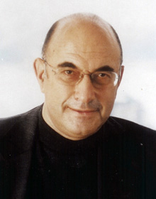
Today, we’re going to move from myth to theory and discuss an argument put forward in the 1980s by Russian-born neuropsychologist and researcher, Elkhonon Goldberg.
We mentioned Elkhonon earlier in the series as the intrepid young researcher who snuck through the USSR’s Iron Curtain and immigrated to the US in the 1970s at the height of the Cold War. For anyone looking to learn more, that history and a detailed account of Elkhonon’s theory can be found in his book The New Executive Brain.
We’ll explain the general outline of his theory, and discuss the key pieces of evidence in favour of it. For today, we’ll go through what Elkhonon’s theory says about differences between the left and right hemispheres. This will set us up for next time, when we cover differences between the frontal and posterior parts of the brain, and address similarities and differences between the sexes.
The key ideas behind Elkhonon’s novelty-routinisation theory of brain function
Before we dive into the details, let’s cut straight to the chase and explain the basics of Elkhonon’s theory. The brain comes in three dimensions: left-right, front-back, top-bottom. All of these dimensions offer interesting ways to look at the brain, but only two of them are incorporated into Elkhonon’s theory: left-right and front-back.

The core of Elkhonon’s theory is that brain functions are organised along a continuum. This continuum refers to the type of information that the brain encounters, ranging from novel stimuli and situations at one end to familiar stimuli, situations and routines at the other. In this way, Elkhonon provides a theory of brain function that accounts for differences in people’s experiences, as what’s novel to one person may be familiar to another, and vice versa.
The novelty end of the continuum is associated with the right hemisphere and the frontal areas of the brain, and the familiar/routine end of the spectrum is associated with the left hemisphere and the posterior parts of the brain. As we alluded to, there’s also evidence of sex differences in how the human brain deals with novel and routine information and situations.
If the distinction between frontal and posterior cortex is unfamiliar, don’t worry, we’ll explain next time when we cover the front-back dimension and sex differences. For now, just know that the brain can be divided into frontal and posterior portions, and that their functions are different but complementary, much like the hemispheres.
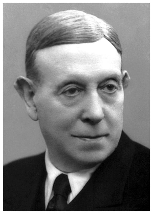
Elkhonon proposed his theory when the field was still bogged down in old ideas which venerated the left hemisphere over the right, and viewed the frontal lobes as expendable liabilities. This view was partly responsible for originating the myths we’ve debunked so far. More darkly, this also led to some of the worst mistakes in neuroscience, such as the practice of frontal lobotomies, which won its inventor a Nobel prize in 1949.
Frontal lobotomies (originally called leucotomies) were later banned when it became clear that surgically removing, silencing or destroying the frontal lobes can have dramatic effects on people’s personality and capacity to live independent lives. The Simpsons poked fun at this tragic history when Ned Flanders became overlord of Earth and forced people to get frontal lobotomies in order to pacify them. To hear it from the horse’s mouth, in The New Executive Brain, Elkhonon writes:
In the old literature the right hemisphere was referred to as the “minor hemisphere” and the frontal lobes as the “silent lobes.” Today we know that these structures are neither minor nor silent, although their functions may be elusive. The functions of the right hemisphere are less transparent than those of the left hemisphere, and the functions of the frontal lobes are less transparent than those of the posterior cortex, precisely because they deal with situations defying easy codification and reduction to an algorithm. So much for the minor hemisphere and the silent lobe! It has taken a longer time to appreciate their functions, but we are beginning to understand their true complexity and the central role they play in our mental processes.
Elkhonon calls his proposal the novelty-routinisation theory. (If you want to get technical about it, as a philosopher of science would, the word ‘theory’ should be reserved for something that’s been accepted by the research community, like Darwin’s theory of evolution or Einstein’s theory of relativity, whereas unaccepted ideas are called hypotheses.) Elkhonon’s theory is not widely known in the field, but he makes an argument that I personally find persuasive enough to share. So, where’s the evidence for all this you ask? Let’s start with the left-right dimension.
Novelty in the right hemisphere, familiarity/routines in the left hemisphere
As Elkhonon said in the quote above, the frontal lobes and right hemisphere have experienced a similar history in parallel. Just as the frontal lobes were once thought to be relatively insignificant, the right hemisphere was often diminished or entirely dismissed by researchers.
For example, people like Michael Gazzaniga once viewed the right hemisphere as the less competent and largely unnecessary back up to the genius of the left hemisphere. At the time, Gazzaniga even called the mental skills of the right hemisphere vastly inferior to those of a chimpanzee, although he doesn’t believe that today.
As we’ve seen in this series, that’s because the right hemisphere is now known to be a crucial member of the cognitive team, as right hemisphere damage can cause a wide range of symptoms and deficits. In fact, research has even shown that the right hemisphere has more to do with IQ scores than the left!
Elkhonon’s theory says that both hemispheres collaborate on all tasks, and are best viewed as a team. However, they differ in their preferences for information and situations that are either novel or familiar/routine. The evidence for this is fascinating and comes from a range of sources.
Lessons from ‘face blindness’ and amusia
For example, some neurological disorders prevent people from recognising certain types of sensory input. Two examples are prosopagnosia, in which people specifically lose the ability to perceive faces (‘face blindness’), and amusia, in which people lose the ability to distinguish pitch and thus can’t perceive music.
Cases vary, but to some people with amusia, listening to music is as unpleasant as hearing a bunch of pots and pans dropped on the floor. Prosopagnosia also varies, with a severe case described by the late neurologist and legendary science writer, Oliver Sacks, in his classic book The Man Who Mistook His Wife for a Hat and Other Clinical Tales.

The key point is that both conditions were thought of as diseases of right hemisphere damage, as this was the cause of prosopagnosia and amusia in most cases. Findings like these helped to establish the idea that creative and emotional stuff are the right hemisphere’s business.
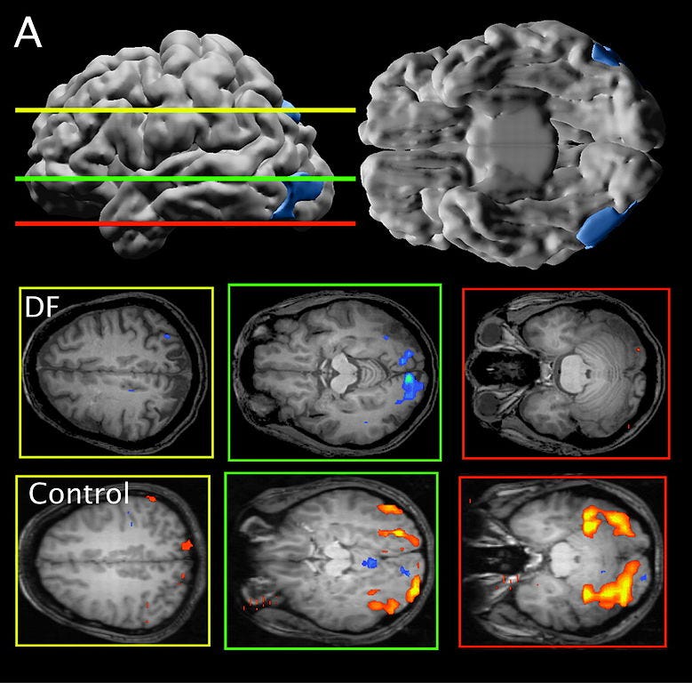
However, further research found that the effect of brain damage depends on whether the faces and music are familiar or novel. Most people are musical novices, and so rely preferentially on their right hemisphere during musical tasks. But expert musicians, to whom music is extremely familiar, rely more on the left hemisphere than the right.
In a similar way, the inability to recognise faces depends on the type of face, as right hemisphere damage prevents recognition of novel faces, but not recognition of famous faces. These findings were published in studies and case reports of people who experienced brain damage (e.g., strokes, head knocks, infections).
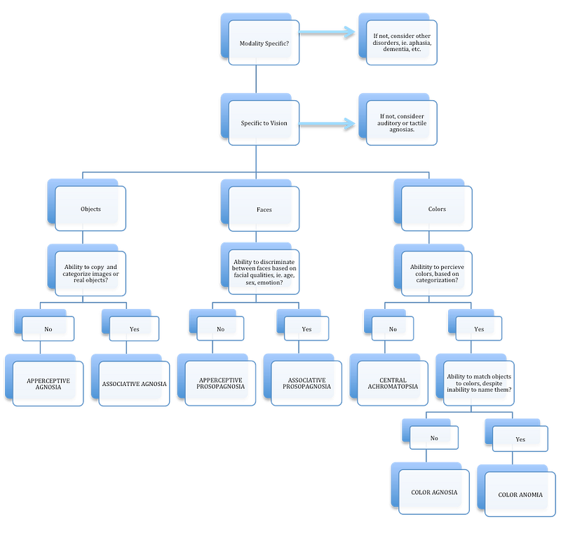
What are ‘agnosias’ and what do they teach us?
Elkhonon also cites other neurological conditions as evidence for his theory. There are two different flavours of a family of conditions known as ‘agnosias’.
Agnosias occur when a person either loses the ability to categorise one or more items (associative agnosias), or loses the ability to identify something (or someone) as an individual entity (apperceptive agnosias). For example, prosopagnosia (face blindness) comes in both kinds, one in which people can’t identify faces (apperceptive), and one in which they can identify individual faces but can’t tell you that they belong to the category ‘faces’ (associative).
People with the apperceptive kind of prosopagnosia can have trouble with everyday things like picking their kids up from daycare. Some report anxiety over the thought that they might accidentally try to pick up someone else’s child, which has happened, though probably not often.
In The New Executive Brain, Elkhonon describes a case of associative agnosia from a patient he worked with as a neuropsychologist. She had a case of visual associative agnosia, where she could see items but couldn’t tell what category they belonged to until she used information from a different sense, like touch. It turned out that this was because a stroke had damaged visual areas of her brain, while the brain regions related to touch remained intact:
[A] middle-aged woman woke up one morning, walked into the bathroom, looked around, saw various objects (a toothbrush, a soap bar, a mirror), and did not know what they were [failure to recall the category they belong to]. But she was able to recognize them [correctly recall their category] when she touched the objects. The woman became scared, got an inkling that something was medically wrong, and had herself driven to a local emergency room. A computerized tomography (CT) scan was performed and a left [hemisphere] stroke was discovered, which must have taken place during the night. This was a typical case of associative agnosia [failure of categorisation] in the visual modality — visual object agnosia. Sometime later she was referred to me for a neuropsychological evaluation and became my patient.
Interestingly, the difference between the failure to identify (apperceptive agnosia) and the failure to categorise (associative agnosia) maps fairly well onto the hemispheres, with failures to identify associated mostly with right hemisphere damage, and failures to categorise associated mostly with left hemisphere damage.
Elkhonon argues that this supports his theory because we use categories (left hemisphere) for things with which we’re familiar, whereas identifying individual members of a category (right hemisphere) inevitably involves novelty, as it means determining what makes something unique.
A shift in left-right hemispheric dominance as we age
Elkhonon also points to an interesting shift in the centre of mental gravity that occurs at around age three. Up until roughly three years of age, the right hemisphere is more active than the left, but this then flips, with the left hemisphere becoming more active than the right. This is at least broadly consistent with Elkhonon’s ideas, as right hemisphere activity declines, and left hemisphere activity increases, as the world becomes more familiar throughout life.
But if that’s true, you could debate whether it makes sense for this shift to occur at around age three. After all, does a three year old really undergo such a major shift in their understanding and mastery of the world? It would also be good to know if this is the case for people like lefties and those with right-brain dominance for language.
Those questions aside, it’s true that a number of observations are consistent with Elkhonon’s theory when you’re looking at a timescale of years to decades. What about minutes to hours?
A brief explanation of brain scanning
This brings us to the world of brain scanning technology. We won’t bother with technical details, but some information about different approaches to brain scanning may be helpful. If you’re not interested in this, feel free to skip to the next section.
MRI (magnetic resonance imaging) uses powerful magnets and sophisticated computer programs to produce a snapshot of brain activity based on the flow of oxygenated blood. When you put a series of these snapshots together, you can reconstruct the flow of oxygen across time, and MRI becomes known as fMRI.
This approach can give fairly detailed information about which areas of the brain are receiving oxygen, presumably for activity. Its weakness is that there’s a limit to how closely most MRIs can look at the brain, and the results lack precise timestamps for activity, although newer technologies are constantly improving on both issues. CT (computerised tomography) scans are similar but use X-rays instead of magnets.
EEG (electroencephalography) involves wearing a cap with electrodes built into it. This gives precise timestamps of when changes occur in your brain’s electrical activity, but not where they occur, as EEG can’t identify which brain cells caused the change in electrical activity. As the EEG cap is worn on the head, EEG recordings are also limited to brain regions that are fairly close to the skull, which excludes much of the brain.
PET (positron emission tomography) involves using a substance that can be traced throughout the brain and body. Researchers use this to assess activity by measuring the consumption of metabolic fuels like the sugar glucose, similar in principle to how MRI measures the flow of oxygenated blood as a proxy for brain activity.
There are other methods and many details, but these are some of the most common approaches to brain scanning. For more on the biology of energy and metabolism, see my other series on cooperation and competition in biology and evolution.
Back to the left-right dimension
So, what about shorter timescales? Do you also see a right-left shift in brain activity over minutes and hours as tasks go from novel to familiar/routine? According to a large number of studies using a range of brain recording techniques and tasks engaging different areas of the brain, the answer appears to be yes.
From remembering word lists and making drawings to perceiving faces/symbols and performing complex behavioural tasks, evidence suggests that the centre of mental gravity undergoes a right-left shift as the task or information goes from novel to familiar/routine. If you want to learn more, you should read Elkhonon’s book (or listen to this talk).
Where does this leave us? In my opinion, Elkhonon marshals a formidable body of evidence that supports his idea that the right hemisphere has a preference for novelty and the left hemisphere has a preference for familiarity/routines. Elkhonon first proposed this theory in 1981, and was a lone voice in the wilderness for a long time.
However, others have begun to jump on board, including an article in Scientific American in 2009. The authors probably should’ve given Elkhonon more credit, but that’s the way it often goes in the world of science, and Elkhonon doesn’t seem salty about it, so I guess no harm done.
Next time
That’s what Elkhonon’s theory says about the left-right dimension of the brain. But as we’ve said, that’s not the end of the story, and Elkhonon also has fascinating ideas about the brain’s front-back dimension.
While recording studies have indeed found evidence of a right-left shift as tasks go from novel to familiar/routine, evidence also suggests that this transition involves a front-back shift as well. Interestingly, however, the frontal cortex doesn’t have the same relationship with novelty as the right hemisphere.
Explaining this difference teaches us a lot about the brain in general, the front-back dimension of the brain in particular, and even leads us to differences between the sexes. We’ll pull on that thread and see what we find next time. Until then!