
AI has shown how little we know about the structure of a cell — we don’t even know half of its components!
Biology textbooks will have to be rewritten. Biochemical analyses have shown that cells contain twice as many structures as previously thought. The discovery is due to artificial intelligence.
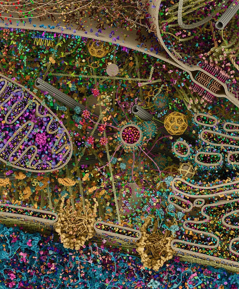
NASA has accustomed us to stunning images of the cosmos. Processed and colored images of distant nebulae and galaxies have always fired the imagination. Some time ago, the agency associated with space helped create an image of one of the smallest objects that surround us — the cells of our body.
Wrongly called a “photograph”, it amazes with its complexity and precise representation of elements such as the nucleus, mitochondria, Golgi apparatus, or endoplasmic reticulum. As it turns out, however, this graphic has even less in common with reality than we thought.
“Scientists have long known that cells have a more complex structure than that described in biology textbooks. Now we have finally been able to look deep into the cells and discover completely new structures” — says Professor Trey Ideker from the Faculty of Medicine at the University of California, San Diego. He is one of the authors of the discovery, details of which were published in the prestigious scientific journal “Nature”.
Not everything in cells can be seen under a microscope
Knowing what our cells are made of is the basis of biology. Disturbances in the work of, for example, cell nuclei or mitochondria lead to serious diseases. That is why cytology, the science of cell structure, is crucial for, among other things, the development of medicine.
Our information on this comes from two main sources. The first is microscopic observations, which most often require killing the cell and staining its components with various chemical compounds. This approach limits the ability to obtain accurate data. In addition, structures smaller than those measuring a micrometer (a millionth of a meter, or a thousandth of a millimeter) are difficult to see through microscopes.
The second source, which allows the study of structures measured in nanometers (billionths of a meter, or millionths of a millimeter), is biochemical research. By using methods such as specific antibodies, scientists can learn about the structure and distribution of those components of a cell that cannot be seen under a microscope. These are most often individual proteins or protein complexes.
Artificial intelligence discovers new structures in cells
“How do you go from micromete to nanometre scale in research? For a long time, biology was unable to cope with this. Only the use of artificial intelligence algorithms helped,” explains Prof. Ideker.
The problem is that research — especially biochemical research — produces a lot of data that humans find difficult to sort through. That’s why researchers at the University of California at San Diego, the Royal Institute of Technology in Stockholm, and Stanford University have entrusted this task to a computer algorithm called MuSIC (Multi-Scale Integrated Cell). It can create a digital model of the cell’s interior by processing available data.
The scientists tested MuSIC on human kidney cells from laboratory culture. The results of the analysis surprised the scientists because they showed how incomplete our knowledge of cytology is.
The algorithm discovered twice as many cell elements as previously described
The MuSIC algorithm found about 70 components in kidney cells. As many as half of them are structures previously unknown to science. Among them was found a protein complex involved in the processing of RNA strands. These molecules play a key role in protein synthesis in cells. They are also used in modern vaccines, such as for COVID-19.
Scientists emphasize that the model created by the algorithm does not describe the spatial structure of the cell. Instead, it indicates the connections between its elements, which is a living organism that can move and change, depending on the situation. Such a map facilitates the understanding of intracellular processes.
The study described in Nature is just a preliminary demonstration of MuSIC’s capabilities. The scientists studied only one cell type and just 661 of the thousands of proteins found in those cells.
“The next step is to carefully go through the entire kidney cell. Then we will study other human cells, cells from different people, and different species of organisms. This will enormously expand our knowledge of cytology,” announces Prof. Ideker.
Source: Nature
Cool that you made it to the end of this article. I will be very pleased if you appreciate the effort of creating it and leave some claps here, or maybe even start following me. Thank you!