
Surfacing issues of peripheral monocyte infiltration in neurodegenerative diseases
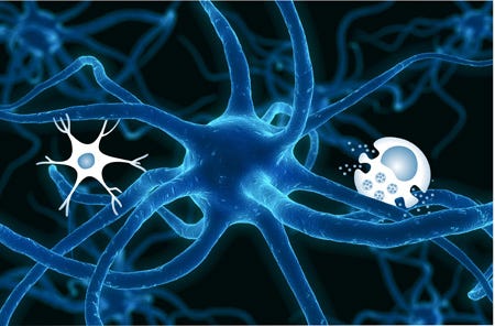
Those of us studying neuroinflammation in degenerative brain diseases, and microglia in specific, are facing an interesting conundrum: what to do with the sneaky bone marrow-derived monocyte. Normally denied access to the brain parenchyma by a resilient blood brain barrier, these blood leukocytes are the precursor to the infiltrating macrophage, which responds to any number of tissue insults, mounting an immune response and cleaning up cellular debris. Do we see them in neurodegenerative diseases and if so, what are they doing?
Until recently, it was thought that peripheral immune cells contributed to the pool of endogenous brain microglia through regular infiltration of the blood brain barrier. This hypothesis was overturned, however, when Ajami et al (2007) demonstrated using a parabiosis mouse model that bone marrow-derived monocytes do not contribute to the population of myeloid cells (i.e. microglia) within the adult brain. Adult microglia are, therefore, a self-sustaining population of cells that we have for the entirety of our lives. The study authors accomplished this by connecting the circulatory system of a wild type mouse with the circulatory system of a mouse whose bone marrow cells glowed green under a fluorescent microscope (in the latter case they simply transferred bone marrow from a mouse where every cell in the body is positive for the green fluorescent protein). After allowing the circulations to mix, the authors looked at the brains of the wild type mice and noticed a complete absence of green fluorescent protein-positive cells suggesting that under normal circumstances, blood monocytes from the bone marrow do not contribute to the brain microglia population. Even after inducing neurodegeneration with an Amyotrophic Lateral Sclerosis (Lou Gehrig’s disease) model in the same parabiotic setup, the authors noted a complete lack of bone marrow-derived cells.
Why does this paper go against the plethora of prior and recent data showing that monocytes do indeed infiltrate the brain during degeneration? Well, it turns out it has to do with the technique of transplanting green fluorescent protein-expressing bone marrow cells, which involves irradiating the entire mouse. And with that, you have an opening of the blood brain barrier. Other papers that do not use irradiation, but other techniques, still note (at least transiently) opening of the blood brain barrier.
Now, it is fair to say that several neurodegenerative diseases do manifest leaky cerebral blood vessels (e.g. Alzheimer’s disease and Multiple Sclerosis) and infiltrating immune cells are thought to effect disease progression in those that do. As you might expect, scientists want to know how to differentiate the infiltrating monocytes from the endogenous brain microglia. Despite similarities between the populations, it seems unlikely that microglia and monocytes function the same in the presence of neurons, given the disparities in developmental origin and milieu, lifespan and reactivity.
In light of the recent data suggestive of a role for peripheral immune cells in neurodegenerative diseases, two camps of thought have surfaced. Camp one, advocates that infiltrating monocytes are bad news for neurons during degeneration. Think of it this way: the partitioning of brain and blood results in unfamiliarity between neurons and monocytes. As a reactive cell type involved in recognizing and eliminating non-self entities, monocytes can become deadly when first encountering neurons and their “foreign” surface antigens. After recognition of these antigens, monocytes are then capable of mounting an intense immune response, secreting neurotoxic quantities of pro-inflammatory cytokines and reactive oxygen species ().
The second camp, on the other hand, believes that infiltrating monocytes offer protection to the neuron — even more so than endogenous microglia. In fact, this camp suggests that infiltration of monocytes into the central nervous system later in degeneration occurs in order to quench the microglial inflammatory response (London et al 2011). There is no doubt that activated microglia in the context of neurodegeneration can play a deleterious role, but how and whether infiltrating monocytes ease microglial activation remains unclear.
Whether good or bad, an important challenge moving forward in the field is differentiating these two cell types from molecular (mRNA expression patterns of specific molecules or receptors) and morphological standpoints. Unfortunately, in order to establish an understanding of their involvement in pathological processes, we need to be able to distinguish them. Although developmentally different, the two populations do share a vast array of receptors and pro- and anti-inflammatory molecules and when each is fully activated, look remarkable similar (resting microglia are intensely ramified with radiating processes, whereas monocytes always look much rounder or ameoboid). Several recent studies have attempted to accomplish this feat and it does seem that there are a few molecules that microglia express, but which are absent in monocytes (Chiu et al. 2013, Hickman et al. 2013, Butovsky et al. 2013). Although this is a good start, more validation will need to be done in the context of disease in order to tease out the roles of each cell type. A huge question in the field at the moment is what happens to peripheral monocytes after they infiltrate the brain parenchyma? Do they remain as monocytes? Die? Or perhaps differentiate into a more microglia-like cell? This, of course, has caused another rift in the field of myeloid cell research and the verdict is still out.
As the broad field of neurodegeneration moves further towards an understanding of disease mechanisms, the immune system’s role is becoming widely recognized over older, more dogmatic hypotheses (e.g. the Amyloid Cascade Hypothesis in Alzheimer’s disease). Now that the initial findings have been established, further study delineating the precise role of each cell type (i.e. microglia and monocytes) in disease onset and progression is required and ongoing. This will inevitably lead to better, more appropriate therapies for neurodegenerative diseases with an inflammatory component.
References
Ajami B, Bennett JL, Krieger C, Tetzlaff W, & Rossi FM (2007). Local self-renewal can sustain CNS microglia maintenance and function throughout adult life. Nature neuroscience, 10 (12), 1538–43 PMID: 18026097
Butovsky O, Jedrychowski MP, Moore CS, Cialic R, Lanser AJ, Gabriely G, Koeglsperger T, Dake B, Wu PM, Doykan CE, Fanek Z, Liu L, Chen Z, Rothstein JD, Ransohoff RM, Gygi SP, Antel JP, & Weiner HL (2014). Identification of a unique TGF-β-dependent molecular and functional signature in microglia. Nature neuroscience, 17 (1), 131–43 PMID: 24316888
Chiu IM, Morimoto ET, Goodarzi H, Liao JT, O’Keeffe S, Phatnani HP, Muratet M, Carroll MC, Levy S, Tavazoie S, Myers RM, & Maniatis T (2013). A neurodegeneration-specific gene-expression signature of acutely isolated microglia from an amyotrophic lateral sclerosis mouse model. Cell reports, 4 (2), 385–401 PMID: 23850290
Hickman SE, Kingery ND, Ohsumi TK, Borowsky ML, Wang LC, Means TK, & El Khoury J (2013). The microglial sensome revealed by direct RNA sequencing. Nature neuroscience, 16 (12), 1896–905 PMID: 24162652
London A, Itskovich E, Benhar I, Kalchenko V, Mack M, Jung S, & Schwartz M (2011). Neuroprotection and progenitor cell renewal in the injured adult murine retina requires healing monocyte-derived macrophages. The Journal of experimental medicine, 208 (1), 23–39 PMID: 21220455
Sennlaub F, Auvynet C, Calippe B, Lavalette S, Poupel L, Hu SJ, Dominguez E, Camelo S, Levy O, Guyon E, Saederup N, Charo IF, Rooijen NV, Nandrot E, Bourges JL, Behar-Cohen F, Sahel JA, Guillonneau X, Raoul W, & Combadiere C (2013). CCR2(+) monocytes infiltrate atrophic lesions in age-related macular disease and mediate photoreceptor degeneration in experimental subretinal inflammation in Cx3cr1 deficient mice. EMBO molecular medicine, 5 (11), 1775–93 PMID: 24142887
Varvel NH, Grathwohl SA, Baumann F, Liebig C, Bosch A, Brawek B, Thal DR, Charo IF, Heppner FL, Aguzzi A, Garaschuk O, Ransohoff RM, & Jucker M (2012). Microglial repopulation model reveals a robust homeostatic process for replacing CNS myeloid cells. Proceedings of the National Academy of Sciences of the United States of America, 109 (44), 18150–5 PMID: 23071306
Originally published at gliosis-zabel.squarespace.com.