
Shots of viruses in augmented reality
Not mere drawings but actual experimental reconstructions. You can view these models in augmented reality with your own smartphone by following the moleculARweb links.
Using our moleculARweb site and new tools to come out soon, we prepared web pages with augmented reality (AR) views of important biological entities (a dedicated story here). Here I share with you various views of viruses that you can launch yourself on your smartphone. Wherever you are, without installing any app, just in your phone’s web browser!
Note that these are not simple drawings or hand-made 3D models, but true 3D reconstructions of viral particles obtained through microscopy techniques of very high resolution -i.e. not using normal optical microscopes but electron microscopes. The 3D models are deposited in the Electron Microscopy Data Base (EMDB) together with the raw data used for their reconstruction. For each example that I show below, I leave you the original EMDB entry and a link to the relevant peer-reviewed publication (highly technical, but just in case you are curious). Besides, I leave you all the links for you to view the viruses yourself in AR.
A giant T4 bacteriophage at the train station
Bacteriophages are viruses that infect bacteria (and archaebacteria). The T4 bacteriophage infects Escherichia coli bacteria. It is among the most studied bacteriophages, in fact standing as a model organism.
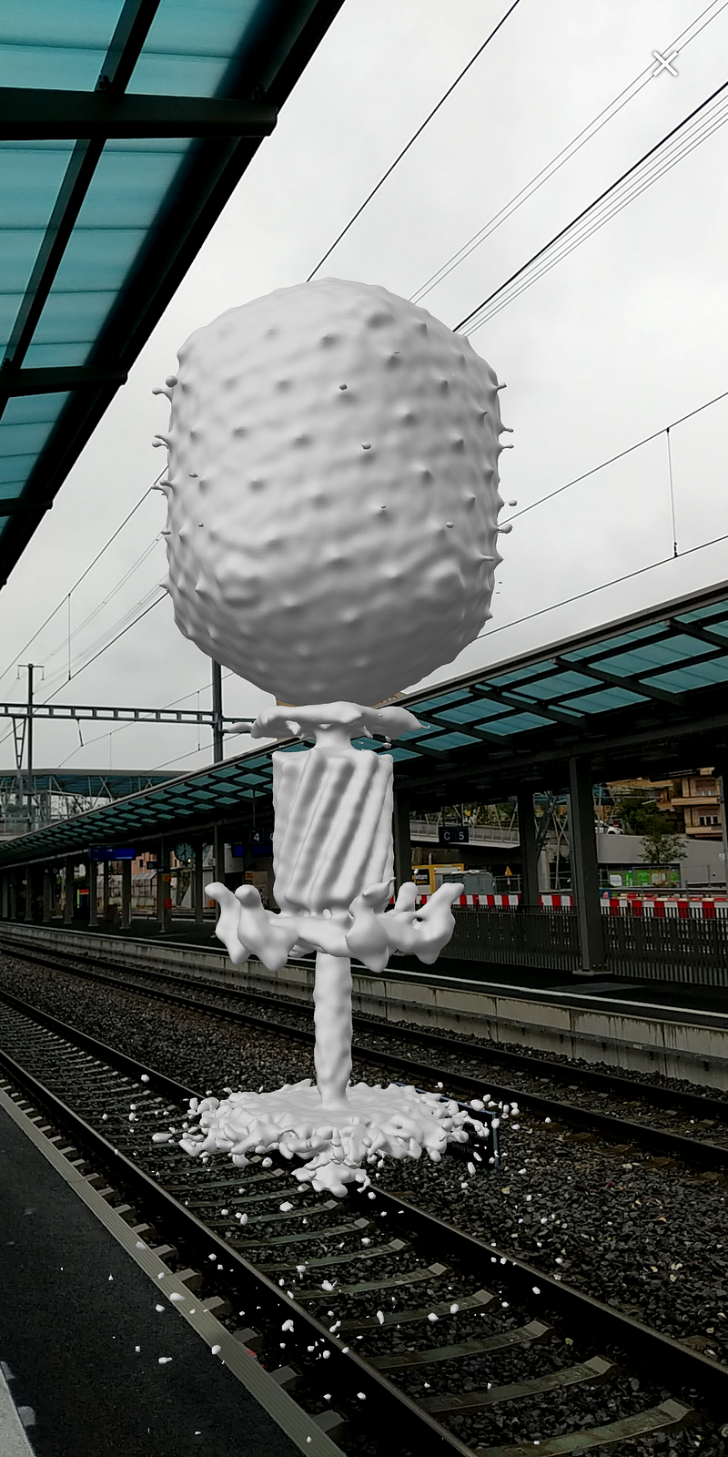
This AR view of the T4 bacteriophage was rendered as an isosurface from a 3D reconstruction based on a cryo-electron tomography map deposited in the EMDB with ID 6082. The associated paper describing it is here (very technical, but in case you are curious). You can see this 3D model yourself in AR by pointing your smartphone’s browser to https://molecularweb.epfl.ch/pdb2ar/t4bacteriophage/
You can appreciate many features of these viruses: the prolate-like capsid, the “drilling machine” and the needle injects the viral genome into the bacterial host. At the end (bottom) of the needle, you see the virus’ base plate fused with a patch of the host’s membrane. What was not resolved in these 3D reconstructions are the “legs” that are usually depicted as sticking out from the base plate -but which are too flexible to be actually resolved in 3D, at least with the current technology.
Another kind of bacteriophage, caught puncturing the two membranes of a Salmonella bacterium
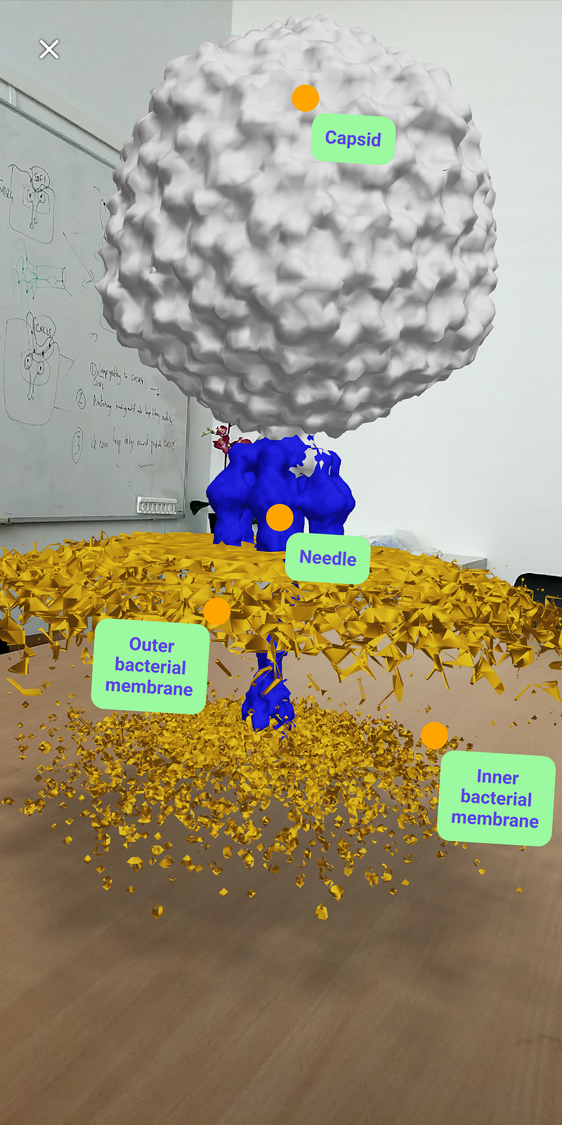
This is a 3D reconstruction based on cryo-electron tomography of another bacteriophage, Salmonella bacteriophage p22 -which, you guessed it, infects Salmonella bacteria. This reconstruction and its associated raw data are deposited in the EMDB with ID 9010. The paper describing this structure is here. To see this one in AR point your phone’s browser to https://molecularweb.epfl.ch/modules/biological-assemblies/virus/virus.html
In this case, the bacteriophage was “caught” when it had punched its needed through the two membranes of a target bacterium. We dissected the 3D map into three main sections: the virus’ capsid (grey, which contains its DNA), the virus’ needle (blue, which injects the DNA into the target cell), and the membranes of the target cell (orange):
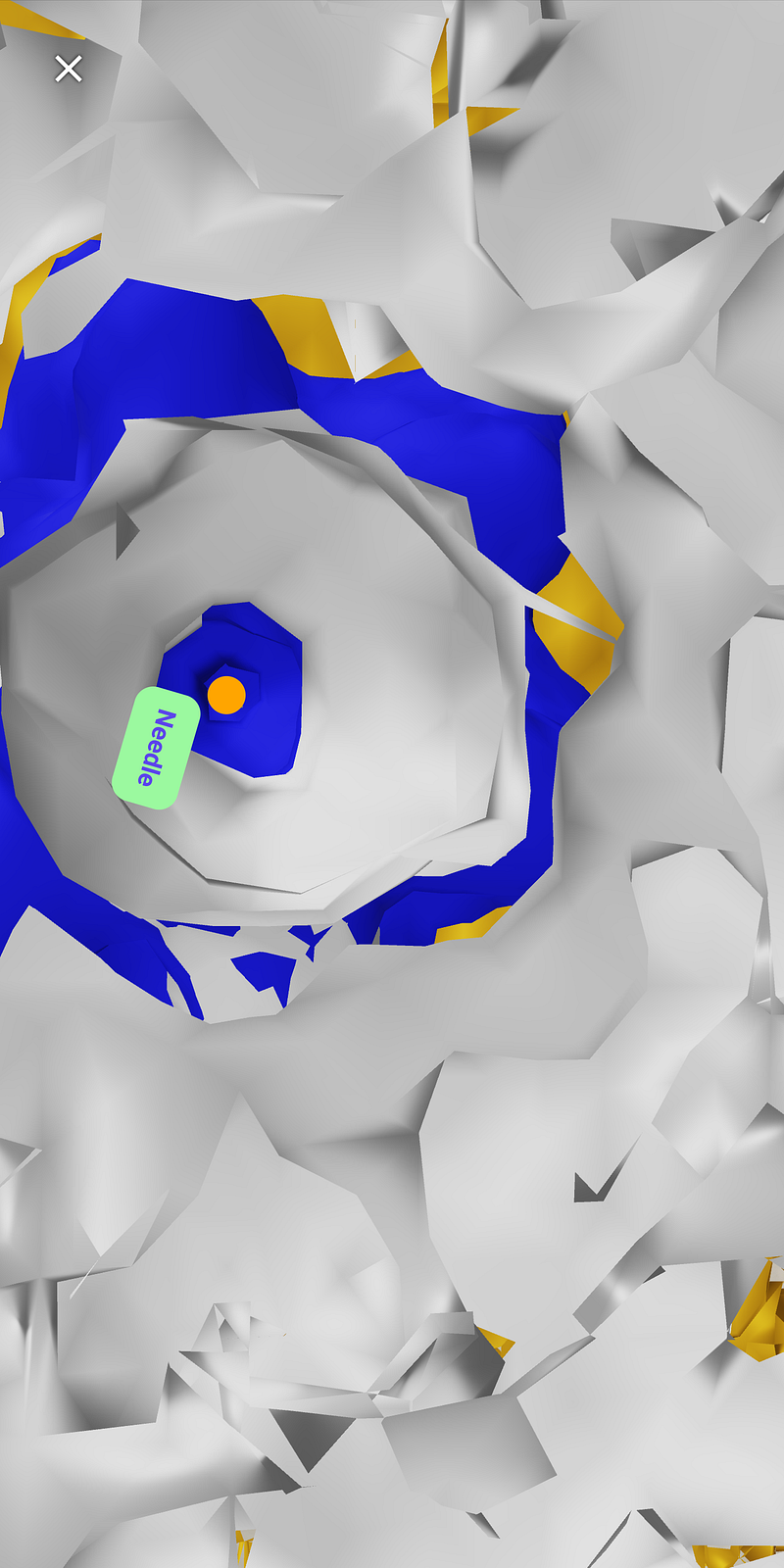
As with all these models, you can move all around it and even get inside. Here for example I moved my smartphone into the capsid and looked down along the needle:
The unfortunate star of the last year-and-a-half: SARS-Cov2
This one doesn’t need much presentation. It’s the animals-infecting virus responsible for the Covid-19 pandemic.
The 3D model below was assembled from cryoelectron tomography and sub-tomogram averaging. It is deposited in the EMDB under ID 30430 and presented in this paper. This model is accessible for view in AR at https://molecularweb.epfl.ch/pdb2ar/sarscov2experimental/.
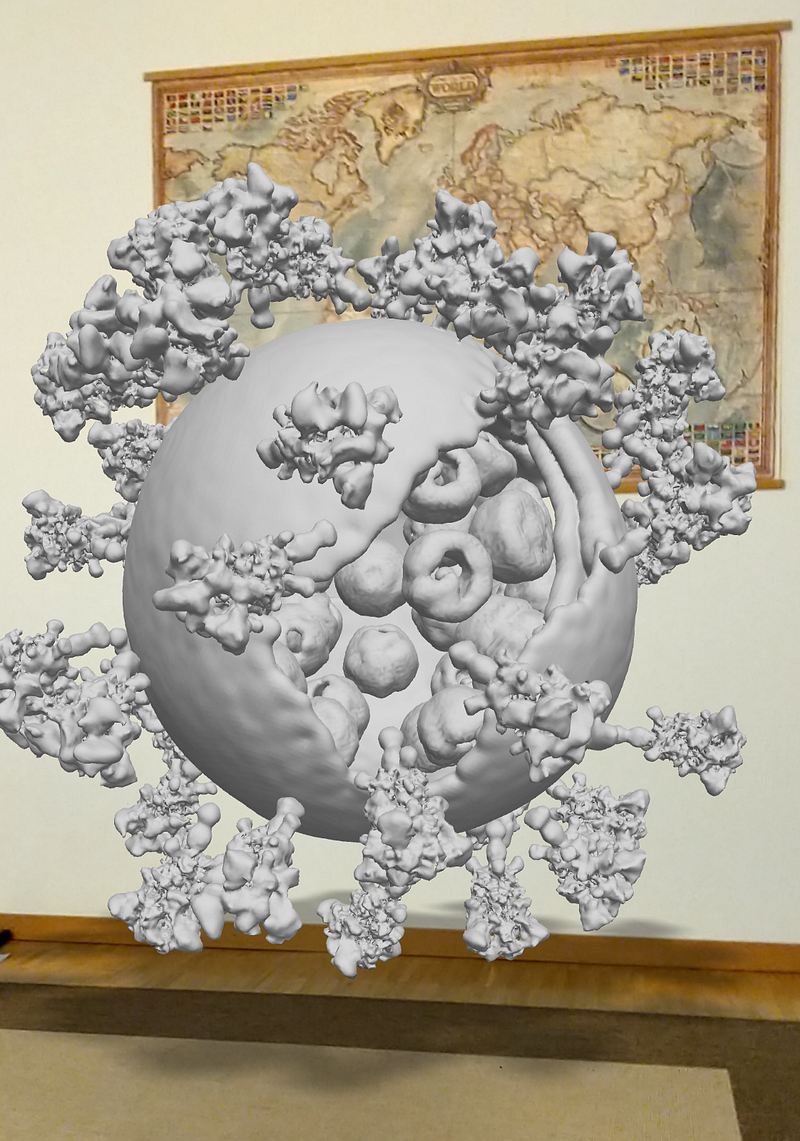
Here it seems to be sketching out a plan to dominate the world (pfff…):
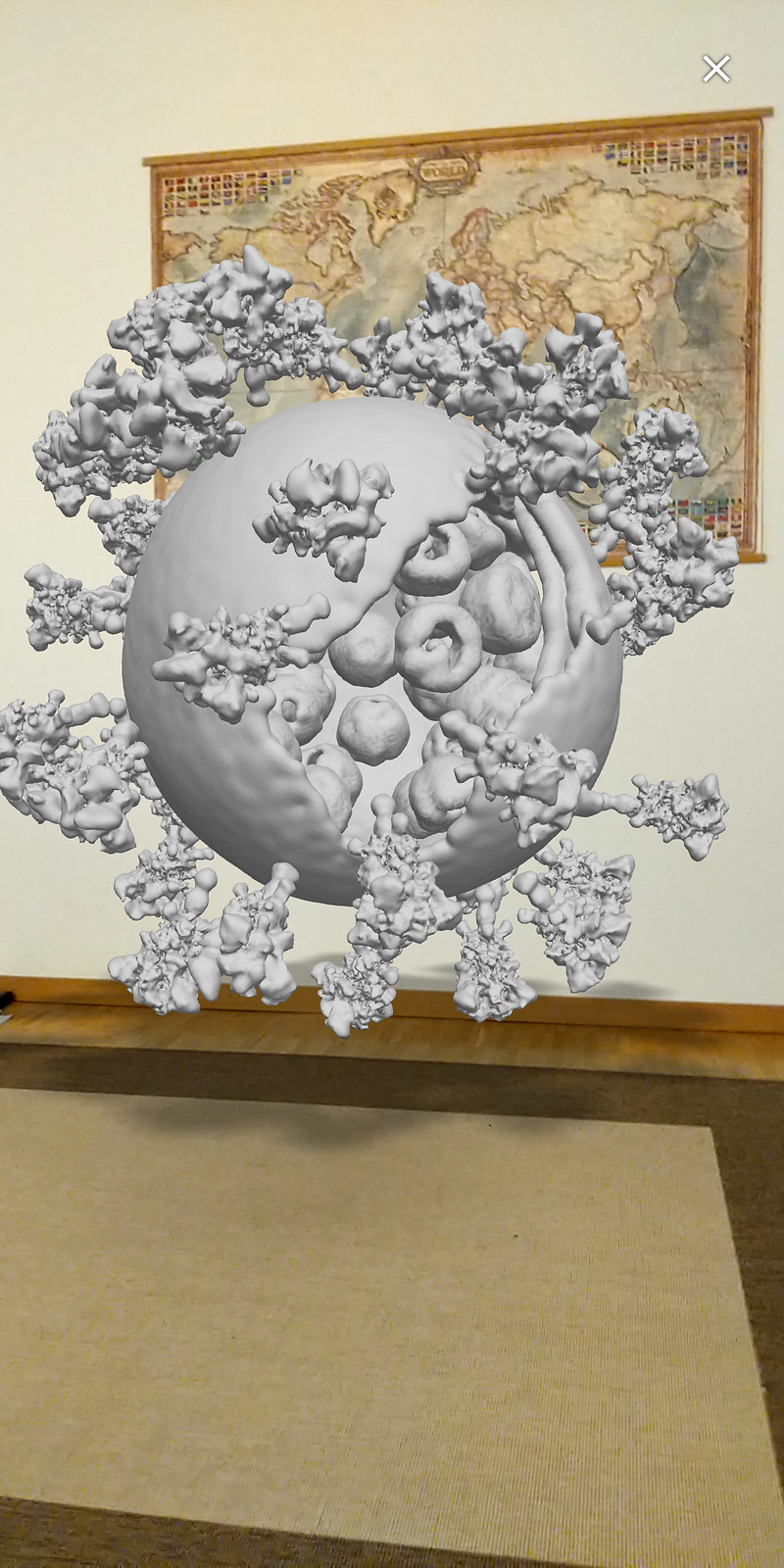
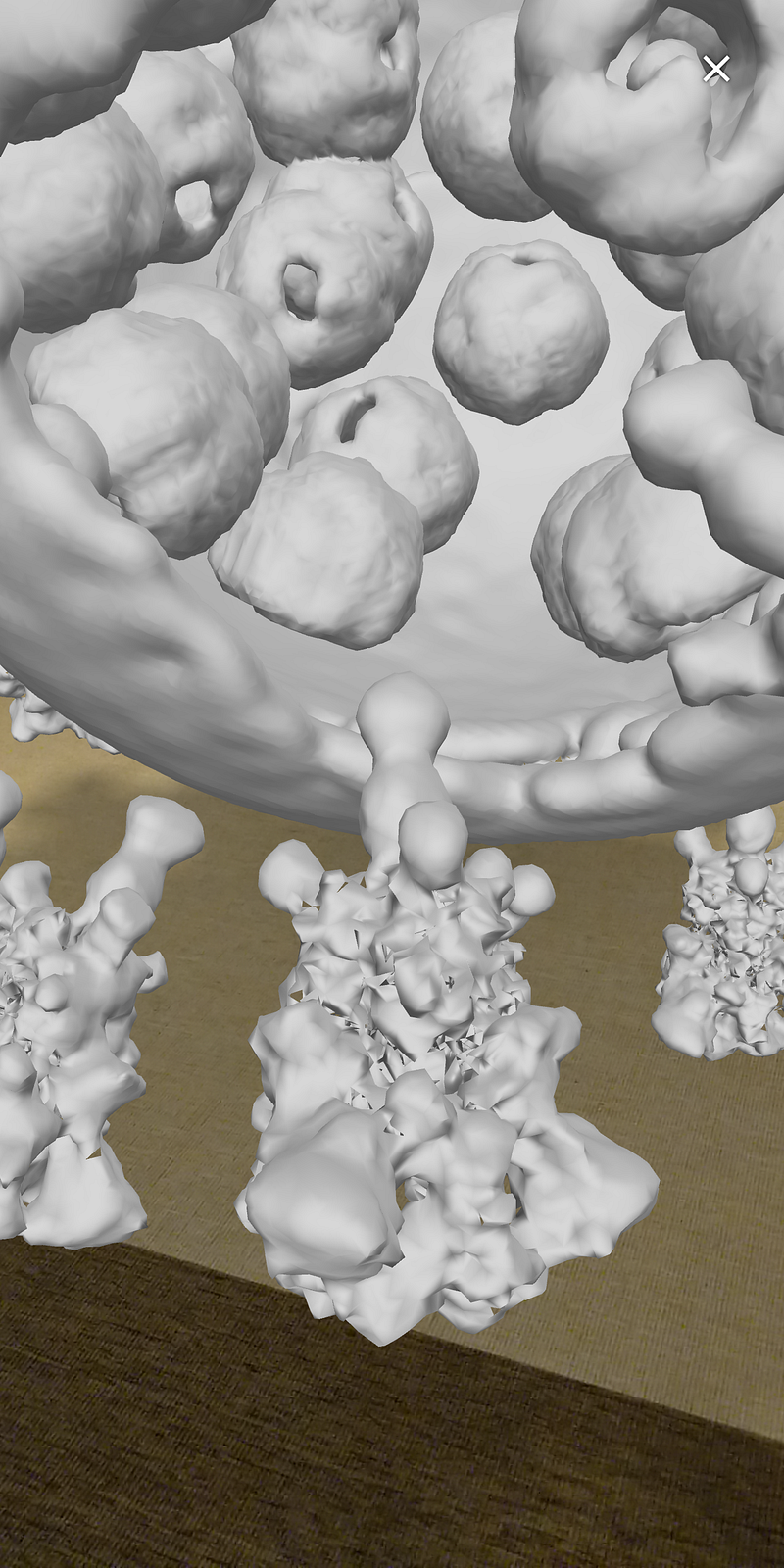
Notice the ribonucleoparticles inside the virus (each ball is an aggregate of proteins and the nucleic acids that contain the genome) and the Spike proteins protruding from the surface:
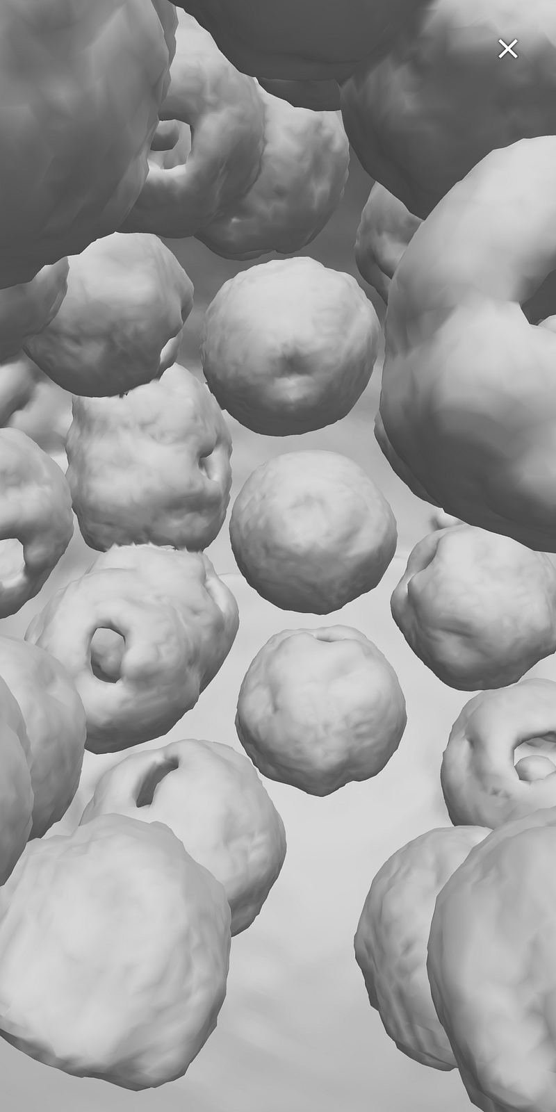
Here’s a view deeper inside, to focus on the ribonucleoparticles:
I hope you can get these AR views to work. Chrome or Firefox on any smartphone should work, but in case of problems drop me a message and I will try to help you. And don’t worry, these won’t infect you!
If the use of web AR was of your interest, check this article on two (free!) AR tools for chemistry and biology education:
I am a nature, science, technology, programming, and DIY enthusiast. Biotechnologist and chemist, in the wet lab and in computers. I write about everything that lies within my broad sphere of interests. Check out my lists for more stories. Become a Medium member to access all stories by me and other writers, and subscribe to get my new stories by email (original affiliate links of the platform).


