
Probing Evolution
How the Placenta, Bearer of Life, Came to Be (With Retrovirus Assistance)
“It just seems like in a way we’re part virus, otherwise we’d be laying eggs,” Coolahan said.

The Placenta
The placenta is a temporary organ that serves as the infant’s lung, food source, and waste removal system. It’s the bearer of life that enable two genetically distinct creatures to co-exist in one body until the other one is born.
“And that, from an immune standpoint, is fascinating, because if you were to receive a piece of someone else and insert that under your skin, that would not last there for three days, your body will actively reject it,” explains Kelsey Coolahan, a medical student at Rowan University.
The co-existence is made possible by the syncytiotrophoblast placental layer — encoded by the syncytin gene —that separates the bloodstream between two the mother and fetus throughout pregnancy. “The wall keeps mom and baby working in harmony and not killing each other,” Coolahan says. “There’s no other structure like this anywhere else in the body.”
Immune-Friendly Cell Fusion: From Env to Syncytin
The placenta first appears in mammals about 130 million years ago. Prior to that, all life forms on earth were laying eggs. This evolutionary transition is facilitated by the syncytin gene that makes syncytin proteins to form the syncytiotrophoblast placental layer.
In 2000, evolutionary bioinformaticians at Genetics Institute, Cambridge, MA, United States, performed a comparative genomic analysis of the syncytin gene. They discovered, for the first time, that the syncytin gene originated from the envelope (env) gene of human endogenous defective retrovirus (HERV-W). As the authors remarked,
“This was a bona fide retroviral envelope protein that had somehow been captured during evolution and been trained to operate in human biology.”
The env gene encodes the virus envelop which is made up of fat, like how our cell membranes are. The virus envelop is what mediate the cell membrane fusion between virus and host cell while suppressing the host immune system — enabling peaceful virus entry.
This membrane fusion and immunosuppression may have been repurposed for how the syncytiotrophoblast placental layer fuses with the uterus lining without provoking the mother’s immune system to destroy it (see figure below). As Joachim Denner, head of Center HIV and Retrovirology Laboratory at Robert Koch Institute, Berlin, said in a concluding statement,
“The fusiogenic and immunosuppressive properties of endogenous retroviruses are evidently the reason we were born.”
Put it in other words,
“Viruses fuse with things in order to infect them,” Coolahan opined. “Now, we get this viral DNA that lets us make a protein that fuses things.”
The Cambridge researchers also showed that inserting this env gene into humans cells endowed the cell the ability to make syncytin protein and to fuse with other cells. Their landmark paper published in Nature was titled:
“Syncytin is a captive retroviral envelope protein involved in human placental morphogenesis.”
[29/3/2020 update; thanks to Chrissie Morris Brady Ph.D.] Syncytin also contributes to muscle repair and regeneration — via myoblast (i.e., muscle cells) fusion — in males only, apparently. Remove this gene and male mice experienced decreased muscle mass. The same was evident in cultured (in petri dish) myoblast of sheep, dog and humans. As the authors of this study articulated,
“These results show a direct contribution of the fusogenic syncytins to myogenesis, with a demonstrated male-dependence of the effect in mice, suggesting that these captured genes could be responsible for the muscle sexual dimorphism observed in placental mammals.”
Sexual dimorphism refers to the distinct characteristics, other than sexual organs, between males and females, such as weight or cognitive processes. This suggests that the syncytin gene might have been repurposed for placental fusion in females and myoblast fusion in males.
Birth Timing: From LTR to THE1B
In 2018, researchers at the University of Cincinnati College of Medicine and National Institutes of Health in the United States unravelled yet another retrovirus element that has chief roles in birth timing.
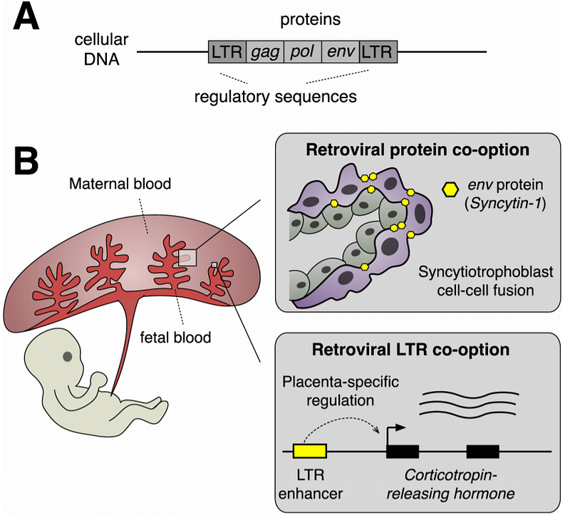
At first, they were interested in how corticotropin-releasing hormone (CRH) — produced in huge amounts in the primate placenta — regulates birth timing. CRH levels increase exponentially as the pregnancy progresses. And misregulation of CRH, thus, may lead result in premature or post-term birth.
The researchers found that CRH expression in the placenta is controlled by a nearby gene sequence, which turns out to be of retroviral origin.
Its name is transposon-like human element 1B (THE1B) that is derived from the long terminal repeat (LTR) of a yet-to-be-identified ancient retrovirus (see figure). Remove this LTR gene and animals cannot produce placenta.
“Their findings implicate an extensive yet understudied role for retroviruses in shaping the evolution of placental gene regulatory network,” says the computational virologist Edward Chuong at the University of Colorado in the United States who was reviewing the 2018 paper.
“…human pregnancy would be very different — perhaps even nonexistent — were it not for eons of retroviral pandemics afflicting our evolutionary ancestors,” Chuong adds.
Retrovirus-Driven Evolution
Retroviruses such as HIV can insert its genetic material into the host genome after it has infected a cell. The host cell then reads this retroviral gene as its own, making proteins that the gene encodes. If the retrovirus infects the genome of the sperm of egg, then the retroviral sequence can be passed on to the genome of the offspring and for generations to come.
For this reason, retroviruses are drivers of evolution. As the professor of evolution and genomics at the University of Oxford, Aris Katzourakis puts it,
“Viral proteins already have functions. It’s much easier to borrow these than to evolve them from scratch.”
The mammalian syncytin and THE1B genes are, therefore, ‘borrowed’ or ‘taken’ from retroviruses. Recent studies, interestingly, suggest that it may not just be mammals after all. Placenta-bearing fishes and lizards have retroviral sequences in their genomes as well.
Another prime example of borrowed function from retroviruses is the Arc gene that is responsible for neuron-to-neuron signalling and neuroplasticity. Speaking broadly, the Arc gene is what drives the evolution of the brain of land animals including humans:
Assimilating functions from retroviruses, however, is a seldom occurrence as optimal functional compatibility is not always guaranteed. About 8% of the human genome are retroviral sequences but the majority are “junk DNA” or “fossilized relics” that are non-functional.
For example, the gag and pol genes of retrovirus— located next to the env gene —have also been integrated into the human genome alongside the env gene (see figure above) but they serve no purpose.
Still, the contribution of retroviruses to the evolution of the brain (in terms of neuroplasticity) and the placenta (in terms of placental immune-friendly fusion with the uterus lining and birth timing) are wonderstruck illustrations of nature at work.


