
How Plants Say Ouch!
One way plant cells talk to each other.
If I get a cut, I say ouch and then maybe suck on the cut to get the blood out of the way so I can see how bad the cut is (eew, gross) or squeeze it to help stop the blood from oozing out. And then if needed, I’ll apply some topical antibiotics and put on a bandaid.
But what caused me to say ouch? Simple.
Nerves at the cut sent a chemo-electric signal to my pain centre and I said ouch!
What happens if a plant gets a cut? Is there a way they can say ouch? And if so, how does it work?
Just like animals and other multicellular organisms, plants do respond to trauma events like dehydration, wounding, pathogen attacks and other insults.
It has been known for more than a century that when a plant is wounded or stressed, it will send out a signal to let nearby cells and other parts of the plant “know” what just happened.
It turns out that one of the signals it sends is waves of charged calcium molecules aka ions.
These waves of calcium ions are very similar to the kind of signalling seen in mammalian nerves but plants don’t have nerves so how is this signalling controlled and propagated?
A number of theories have been put forth to explain how plants might do this but none of them had been rigorously proven to be true.
A recent paper published in Science Advances by Bellandi and others from the Crop Genetics group at the John Innes Centre in Australia reveals that this cascade of ionic calcium waves is actually initiated and modulated by an amino acid called glutamate.
In this article, you’ll learn how glutamate triggers the waves of calcium ions that communicate the wound and thereby help the plant respond to being wounded.
It’s how plants say ouch.
What did we know?
The study we’re looking at extends the work of researchers from the late 19th and early 20th centuries who looked at how plants respond to wounding and stress.
What these studies showed was that plants responded to various kinds of stimuli such as attacks by pathogens or other herbivores, osmotic stress and wounding. But the molecular details were still being figured out.
And because there are different stimuli, different hypotheses were proposed to explain how each might work.
Not surprisingly, one of the fastest and most dramatic responses took place when a plant was wounded.
What the early researchers detected when the plant was wounded was a wave of calcium ions.
Not only was the calcium ion signal given to adjacent cells but it was transmitted throughout the plant and could even be transmitted through dead tissue!
How did this happen?
Remember, plants are highly complex organisms and similar to animals, are made up of many different kinds of tissues and cells. And they have a vascular system composed of two components; the xylem and phloem.
The xylem forms a continuous channel made from cells that are no longer alive. This channel is one of the ways that nutrients and other chemicals reach distant parts of the plant.
The phloem transports food and other nutrients including sugar and amino acids from leaves to storage organs and growing parts of the plant.
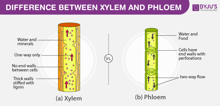
One important feature that distinguishes the two types of vessels is that the xylem generally moves water and minerals in one direction only; from the roots to the rest of the plant. In the phloem, the nutrients, sugar, amino acids and other substances can move in both directions, up from the roots to the above-ground parts or from the above-ground parts to the roots.
Given that the vascular system reaches throughout the plant and extends into all its tissues, maybe plants used it to signal trauma.
Back to the calcium waves.
Waves
Ok, so a plant has been wounded and a calcium wave has been generated.
What exactly is a calcium wave? Is it like the wave we see on the beach or something different?
Quick answer; it’s kinda like the waves you see on the beach but there are differences. Think about the kind of waves that you see on somebody’s heartbeat monitor in the hospital. You see a wavy line with a high point that is generated every time the heart beats.
Here’s a typical mathematical wave diagram where “y” shows the concentration at a given time “t”.
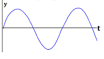
If you measure the concentration of calcium ions at and nearby the wound site, you will see a similar picture generated, where the concentration of calcium gets higher then lower then higher again just like the heartbeats. The peaks are the waves of calcium ions.
Calcium ions are stored in plant cells in specialized little organelles.
In plants, calcium waves don’t just occur spontaneously. They show up during these trauma events.
So what is causing these waves of calcium in plants to occur?
Various theories suggested how these waves might be transmitted. Maybe it travelled along the cell membranes? Maybe some kind of pressure wave formed and flowed through the xylem? Or phloem?
These theories were certainly possible but they lacked one essential thing. They failed to explain how waves were transmitted from one cell to the next and so on.
And that’s what Bellandi’s research looked at; how were the waves transmitted?
To answer that we need to know more about calcium ions. Both plant and animal cells most commonly use calcium ions as what biologists call 2nd messengers.
But if they are a 2nd messenger, that means that their appearance is controlled or triggered by a primary messenger. So what primary messenger were the plants using to stimulate and produce the calcium waves? And how might they do that?
And where were these signals being sent? How did they get from cell to cell?
Questions, questions and more questions!
A New Theory
Dr. Bellandi in Dr. Faulkner’s lab in the Crop Genetics Group had an idea.
Faulkner’s group studies structures that form channels or “bridges” between plant cells. These bridges are called plasmodesmata. For the most part, they are located in the living phloem cells.
Plasmodesmata were first reported by a famous botanist, Eduard Strasburger, in 1901 and are only found in the cells of plants and algae. In higher plants, every cell is connected to its adjacent cell by these channels. In typical higher plant cells, the number of plasmodesmata in each cell can range from 1,000 to 100,000!
That’s a lot of connections.
Here’s a nice graphic image taken from here to help you visualize plasmodesmata.
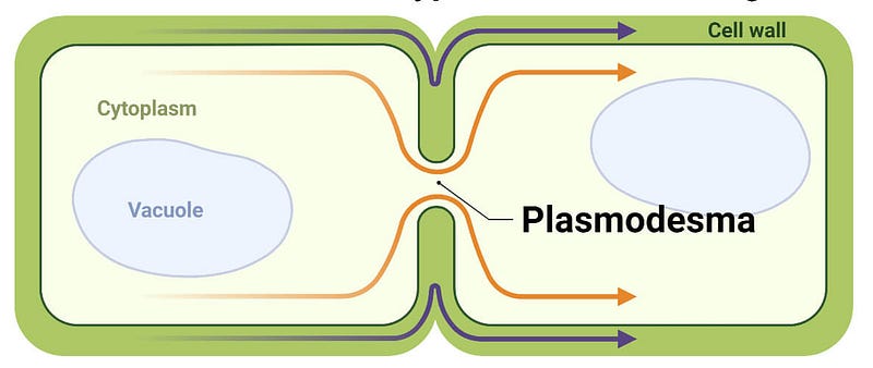
You see two cells encompassed within a cell wall (the wide green band) and a single plasmodesma forming a conduit or channel that connects them. It’s important to emphasize that this image is not even close to being drawn to scale and in a real plant, there would be hundreds or thousands of these plasmodesmata connecting these two cells.
Because water, fluid, proteins, small RNAs, hormones, and metabolites are transported through them during developmental and defence signalling, plasmodesmata are essential for plant life.
One possibility the group looked at was if the messenger signals travelled through the cells via the cytoplasm by using plasmodesmata.
How could they test this idea?
The Research
They decided to use Arabidopsis thaliana, a plant that is often used to investigate cellular biology and genetic and molecular pathways in plants.
A. thaliana grows all over the world as a weed and is in the Brassicaceae, the crucifer, mustard or cabbage family which includes cabbage, kale, cauliflower, broccoli, collards, turnip, Chinese cabbage, rapeseed, common radish and horseradish to name a few common members! You can eat it in a salad or cooked, but because of its small size, it is not highly sought after.
Here’s a full-grown plant. It’s less than 30 cm (12 inches) tall.
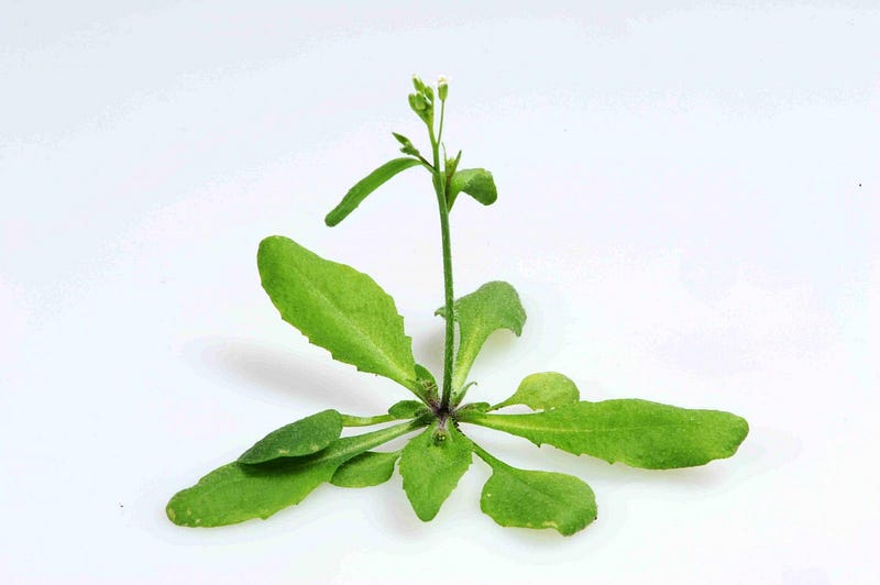
There are many reasons for its use in laboratory plant sciences including its small size, life cycle that is completed in about 6 weeks, small genome, and a single plant can produce several thousand seeds.
Because A. thaliana is such a popular model organism there are thousands of different genetic strains and it is pretty straightforward to create your own using modern genetic engineering techniques.
For their experiments to test this hypothesis, they created strains that contained a calcium reporter located throughout the plant. The reporter fluoresced a particular colour whenever it bound a calcium ion.
If you have a device that can monitor the appearance of this colour, you can track the fluorescence.
Detecting fluorescence
I’d like to spend a few minutes describing exactly how this whole reporter system works because it’s pretty cool!
First, you need to know about tiny pieces of DNA called plasmids.
Here’s how the American National Human Genome Research Institute defines plasmids:
“A plasmid is a small circular DNA molecule found in bacteria and some other microscopic organisms. Plasmids are physically separate from chromosomal DNA and replicate independently. They typically have a small number of genes — notably, some associated with antibiotic resistance — and can be passed from one cell to another. Scientists use recombinant DNA methods to splice genes that they want to study into a plasmid. When the plasmid copies itself, it also makes copies of the inserted gene.”
So we have these little circles of DNA that are found in bacterial cells, make copies of themselves independently of the chromosomal DNA and are one of the primary tools that molecular biologists use to study how genes work.
If you look at the figure below, you can see where they are normally found in the bacterial cell.
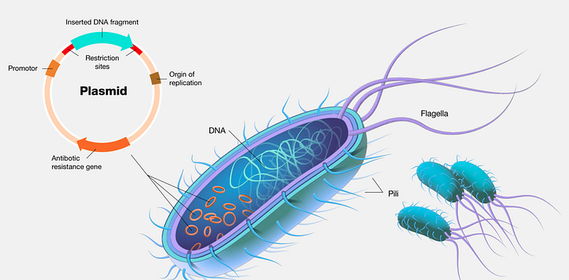
One of the reasons plasmids are such an important tool is that we have figured out ways to isolate and purify them from bacterial cells so that we can alter their DNA or insert other pieces of DNA into them. Then we sequence them to confirm the alteration was what we intended.
You can also splice pieces of DNA into them. These pieces of DNA are either sequences you want to study or they specify a protein you want to investigate. When the plasmid makes a copy of itself, the gene you inserted comes along for the ride and if it encodes a protein, the cells containing the plasmid will make that protein.
Another important characteristic is that plasmids often contain genes that confer antibiotic resistance to bacteria. That’s how superbugs like methicillin-resistant Staphylococcus aureus bacteria — commonly called MRSA — acquire and maintain resistance to so many antibiotics.
So what’s the point of all this info?
The beauty is that you can use a combination of these features to select plasmids that contain the gene fragments you have inserted into them. This is called molecular cloning and the plasmids that are generated are usually referred to as constructs. (TMI, I know, but what the heck!)
Plasmid constructs that researchers insert into foreign host cells are often called vectors because they carry foreign DNA into the host cell.
The 1970s was when plasmids were first designed as vectors for delivering custom pieces of DNA inside a host cell and kicked off the modern era of molecular biology and genetic engineering.
Nowadays, there are lots of companies that sell kits to help you make the plasmid you’re interested in creating so it turns out that there are at least a gazillion plasmids out there that researchers have made to help them investigate the genes and other DNA sequences they are interested in.
Bottom line: we can use plasmids to study a molecular phenomenon of interest.
And that’s what Bellandi and colleagues did.
Ok, back to our study of calcium waves in wounded plants.
In these experiments, Bellandi and colleagues obtained two reporter plasmid constructs designed and made in another laboratory; one detected calcium ions and the other one detected the amino acid glutamate. When either of these constructs was present in a given plant if the target was detected, the plasmid would fluoresce distinct colours or wavelengths of light.
If you want to know specifically how this works, here’s the deets from Dr. Faulkner.
The constructs that detect calcium and glutamate are engineered fluorescent proteins (derived from green fluorescent protein from jellyfish). The proteins have been split in half and rearranged (flipped) so that their head and tail are now in the middle of the protein with a calcium or glutamate protein inserted between them. These are circularly permuted fluorescent proteins. When that middle protein binds calcium (or glutamate if it’s the glutamate variety) it flips the two halves of the protein back together so that they make a functional fluorescent protein. Therefore, without the calcium, the protein doesn’t accept and emit photons in the range we are activating/detecting, but when the calcium is bound it does.
That was quite a mouthful, eh?
Here’s my plain language version:
Using strains of A. thaliana that contained these plasmids, they were able to detect the fluorescence when calcium or glutamate was released and bound the reporter constructs.
They took pictures of this fluorescence prior to, during and after wounding to detect calcium and glutamate.
So what did they see?
Results
Now that you know how this all works, let’s see what they did to test their hypothesis about how the calcium waves are propagated when communicating distress from a wound site to the rest of the plant.
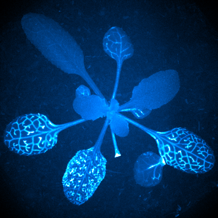
First, let’s take another look at our article’s feature image.
If you look closely, you can see the wound — the stem with the bright white tip at about 5 o’clock- and the leaf that was severed is below it to the right. You can also see that the three leaves still attached to the plant are also brightly fluorescing with very similar and specific patterns. Two others are also fluorescing, but not as vividly.
What the fluorescent patterns are actually showing is the vascular phloem tissue.
But is this pattern always present or only seen after wounding?
And here’s your answer to that!
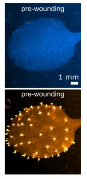
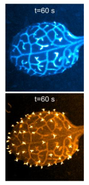
The pictures above show images of calcium-generated fluorescence in the blue images on top and glutamate-generated fluorescence in the brownish images below. They are all of the same leaf and were taken before and then one minute after wounding.
As you can see, the patterns of calcium and glutamate fluorescence one minute after wounding overlap and are nearly identical in their relative levels of brightness!
Pretty cool, eh?!
In addition, they were able to determine that the glutamate wave preceded the calcium wave and was in fact, the primary messenger that triggered it.
What Does it All Mean?
In this article, I only told you about one part of their research, the primary and second messengers of glutamate and calcium during plant wounding. They did additional work so feel free to read the whole article if you’re interested.
Remember, Dr. Faulkner’s lab is especially interested in learning about the roles plasmodesmata play in plants.
So…..
What role did the plasmodesmata play in all of this?
As a result of this work, they were able to rule out several of the former theories about how and when calcium waves were propagated:
The fluorescence showed the calcium waves were not initiated by a pressure wave in the xylem which thereby ruled out the “squeezed cell hypothesis” as the waves did not move through the cytoplasm via plasmodesmata.
That means the waves moved through the cell wall complex.
So plasmodesmata were not involved in communicating signals about being wounded.
This led them to the conclusion that “a century-old model of a xylem-mobile Ricca factor applies to this response” and that transmission of calcium waves can be explained by the passage of glutamate through the cell wall complex using the xylem as it is continuous with it.
Dr. Faulkner explains it this way; “The glutamate and calcium waves are connected — glutamate triggers the calcium response. You could imagine it with an analogy of a corridor. The glutamate rushes down the corridor and as it passes a door it kicks it open. The calcium response is the door opening. Up to now the assumption has been that what moved down the corridor was hydraulic pressure or a series of propagating chemical reactions, but our study shows that this is not the case.”
As you saw in the feature image, not all the plant’s leaves showed the fluorescent calcium wave. They suggest that this is probably due to the amount of glutamate produced at the wound site. As it moves down the “corridor” opening the calcium “doors”, it is gradually used up and at some point, the concentration is not enough to trigger any more calcium releases.
It is important to note that while this calcium response was definitely important in the initial response to wounding, the entire plant response or what is called systemic response requires many more signals to actually close and “heal” the wound.
So it is likely that calcium waves do not initiate all systemic defence responses.
And a bit more from first author Bellandi:
“We’ve shown that calcium waves are synchronous with glutamate waves, and their dynamics match transmission by diffusion and flow. This research makes us rethink what we know. I am hoping that our research will inspire debate and allow people to take a fresh perspective on data which has been in the field for a long time.”
Another important benefit of this research is that they developed quantitative, live imaging methods that can measure the dynamics of local, vascular, and distal calcium waves with high resolution.
Using these methods, they were able to use fluorescence to track and measure the amount of an individual molecule as it travels through the plant.
That’s remarkable!
Using this technique they were able to profile and quantify the dynamics of glutamate and calcium waves they saw in response to wounding the plants.
So not only did they answer a question. They also developed a method that they and other researchers can use when doing this kind of research.
On top of all that, you got to learn or be reminded about higher plant anatomy and molecular biology, plasmids, DNA and fluorescence.
And this is one of the things I really love about science. You don’t know where or when someone is going to come along and tell us about something really fascinating that has been brewing in the literature for decades or more.
In this case, the question had been lingering for over a century!
Until next time,
Rich
Sources
- Diffusion and bulk flow of amino acids mediate calcium waves in plants by A. Bellandi, et al., in Science Advances, (Oct 2022)
- The science of how plants register trauma catches a new wave, John Innes Centre press release, (Oct 2022)
- Red fluorescent genetically encoded Ca2+ indicators for use in mitochondria and endoplasmic reticulum by Jiahui Wu, et al., in BiochemJ, (Nov 2014)
- Wikipedia Arabidopsis page.
- Wikipedia 2nd Messengers page.
- Wikipedia Plasmodesma page.
- The Biology Notes Plasmodesmata page.
- personal communications with Dr. Christine Faulkner.
If you’d like to get Updates from Biology4Everone, you can sign up here. The updates also include short pieces about naturalist topics and what I’m reading. They come out every few weeks so you won’t have your inbox flooded with emails from me, I promise!


