
Did Algae Gain Eyes to Hunt With?
How cellular munchies led to bioluminescent, photosynthetic, predatory algae with eyes
1. Let’s eat…
When the first cells on Earth began to eat other cells over a billion years ago, a whole new world opened up. Before this dramatic new innovation in cell biology, the vast majority of single-celled life for the first few billion years simply gathered free energy from the sun (we call them phototrophs) or chemical energy (chemotroph) from the depths of the Earth itself. But the novel act of a cell eating another cell (phagotroph), like you or me chowing down on a steak, literally rewrote the books on Evolution here on Earth.
Today, we’re educated early to the process of cells engulfing other cells — since elementary school, we’ve seen amoebas eating paramecia or neutrophils in our blood engulfing bacteria, “nature, red in tooth and claw” on a microscopic scale.
This process of cells of hunting and eating other cells laid the foundation for complexity of the plant and animal kingdom that we see today (including our insanely complex immune system — see the neutrophil video linked above). But we see a tremendous level of complexity even within single-celled organisms as well, rooted in that ancient act of eating.
We’ll explore one of the best examples of what amazing creations evolution has wrought, the single-celled dinoflagellate, and how cells eating cells gave rise to this complexity — and to ours.
2. Peepers on plants that hunt…
Dinoflagellates are freakin’ amazing. They are diverse, microscopic, single-celled algae. We know about them because some cause red tides, poisoning shellfish beds, and sometimes the people who eat the tainted mollusks. They are one of the causes of ocean bioluminescence, where at night bright, fluorescent blue-green waves crash to shore, and kayakers or surfers stir up glowing tracks in the water.
Many dinoflagellates have spiky armor plates, and all are mobile, using whip-like cords called flagella from which they get their name. Many are photosynthetic, able to convert sunlight, water, and carbon dioxide into chemical energy stored as sugars. And many of these algae are predatory instead of photosynthetic. But on top of the amazing skillsets of these luminescent, mobile, photosynthetic, predatory algae, a special group of these dinoflagellates has peepers. Eyes.
This special group is a tiny and rare family of dinoflagellates called the Warnowiaceae, and are among the most complex single-celled organisms we know of. They are both photosynthetic and predatory, probably consuming other dinoflagellates and planktonic creatures.
Warnowiids (the name for members of the Warnowiaceae family) employ a range of complex sub-cellular machines such as:
- nematocysts — which throw out prey-capturing filaments
- trichocysts — which extrude threads in response to stimuli
- pistons — which rapidly extend and contract a long rod that may be used for locomotion or to capture prey
- ocelloids — the eye of the algae
3. Ocelloids are like eyes…
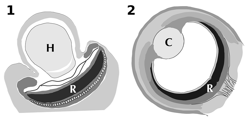
Ocelloids are subcellular structures in Warnowiid dinoflagellates and have all the components we associate with camera-like eyes in vertebrates like us, and in cephalopods like squid and octopus:
- cornea — the clear outer layer of the eye which gathers light
- iris — rings which limit light like the diaphragm shutter of a camera
- lens — focuses incoming light
- retina — receives the focused light and generates a chemical signal.
Of course, one of the first questions we want to ask is: what do these eyes see?
The answer is we don’t know. Warnowiids are rare, difficult to keep alive when captured, and so far, impossible to raise in culture. So, it is very hard to test the creatures, to determine whether these microscopic eyes function and what the Warnowiid sees.
Limited tests to date suggest that these complex organelles are light-sensitive, are damaged by strong light, and contain proteins called rhodopsins that are associated with vision in all the eyes we know of. Other circumstantial data show that Warnowiids contain digested remnants of other dinoflagellates, suggesting that they are hunters and perhaps use their eyes to capture prey.
Of course, we know of microscopic hunters that do not need eyes to chase their prey, using chemical cues instead. So the question remains: why do Warnowiids need such complex eye-like structures?
4. How to build ocelloids…
Even more fascinating than knowing a single-celled photosynthetic alga has an eye, is how it is built. For our eye, we have a pretty good understanding of the cells and proteins making up the different optical structures. For example, we know our lens is made of a single layer of epithelial cells on the surface and the bulk is a transparent protein called crystallin; we know the retina is made of cone and rod cells packed with light-sensitive rhodopsin proteins which detect visible-light photons.
The sophisticated camera-like eye in Warnowiids cannot be made of cells, since the entire organism is a single cell. So how is its eye made?
Two papers, one by Hayakawa et al., published in 2014 in the journal PLOS ONE, and another by Gavelis et al. in 2015 in the journal Nature, help us understand how these ocelloids are built.
It turns out Warnowiids have repurposed several well-known sub-cellular components into the service of vision.
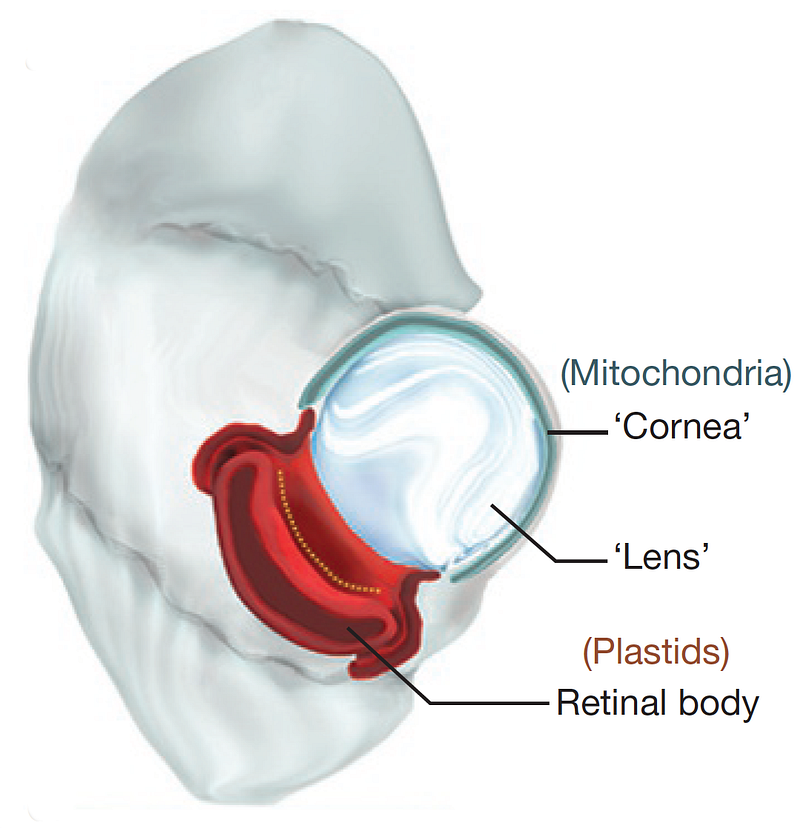
For example, all eukaryotic cells (cells with a nucleus) have mitochondria, the energy-producing organelle within each cell. In the Warnowiids, some mitochondria are drafted into making the equivalent of the light-gathering cornea in the ocelloid.
The lens-like structure in the ocelloid is made from a common cellular structure called a vesicle — which is a membrane-bound sac.
The iris-like structure in an ocelloid is formed from something called a thylakoid — a flattened pigmented sac where the light reactions of photosynthesis occur.
And the all-important retina, the structure in our eye that receives and processes light into chemical signals, in Warnowiids is made of a photosynthetic organelle called a plastid. The photosynthesizing chloroplast in all the green plants we are familiar with is an example of a plastid.
Of all the components in the Warnowiid cell making the ocelloid, the mitochondria and plastids are the most interesting. They are endosymbionts — once free-living bacteria-like organisms that were engulfed by a host cell and passed on to their progeny.
5. Endosymbionts — you will be assimilated…
One of the most controversial and revolutionary biological concepts was the idea that major evolutionary leaps can occur by a process called endosymbiosis.
In 1967, Lynn Margulis was an adjunct assistant professor at Boston University. She had been married to Carl Sagan between 1957 to 1964, and in 1967 married physicist and crystallographer Thomas Margulis. 1967 was also the year her highly controversial paper titled “On the Origin of Mitosing Cells” was published in The Journal of Theoretical Biology, after being rejected by fifteen other scientific journals (the paper was attributed to Lynn Sagan, her name when the journal first received her paper).
But publication of her opus on endosymbiosis was just the beginning of a lonely multi-decade long campaign for her ideas to not only be accepted, but even heard.
In the 1967 paper, Margulis argued that within “higher” eukaryotic cells some critically important organelles were once free-living prokaryotes (cells without a nucleus like bacteria). These free-living cells were symbiotically engulfed by other cells to eventually become essential parts of the complex eukaryotic cell.
You may be familiar with the mitochondria, the power-plant of all eukaryotic cells from yeast to human. Margulis’s hypothesis said that billions of years ago, very early in the evolution of life, photosynthesis developed to capture the energy of sunlight. A byproduct of photosynthesis is oxygen which was toxic to most life at the time. Soon, photosynthesizers produced so much oxygen, they created an oxygen crisis.
Aerobic (oxygen-using) prokaryotes evolved to survive in the new oxygen-laden environment. Some anaerobic (oxygen-avoiding) prokaryotes learned to survive by ingesting aerobic cells and figured out how to co-operate. The aerobic cells became our mitochondria. Eventually, this symbiotic relationship became essential to survival. The term biologists use is obligate endosymbiosis.
The acquisition of endosymbiotic mitochondria was the first step to a more complex cell with a nucleus: the eukaryote.
The other classic endosymbionts Margulis wrote about are less discussed now, but as essential to life today. After the oxygen crisis, some of the newly evolved eukaryotes engulfed one or more prokaryotic photosynthesizers which became obligate endosymbionts known as plastids. One of their descendants are the chloroplasts which we find in all higher plants today.
A third example is not so critical to our story of ocelloids, but is interesting and related nonetheless. Spiral, undulating cells called spirochetes (like those today which cause syphilis and Lyme disease) were engulfed by eukaryotes and became the motor driving their mobility. That motor is now the flagella, the whip-like driver of cells like our bioluminescent dinoflagellates.
Lynn Margulis died in November, 2011 of a stroke. Her hypothesis had finally been experimentally confirmed in an important 1978 paper by R.M. Schwartz and M.O. Dayhoff. Since then her endosymbiosis theory gradually and eventually became accepted.
British biologist Richard Dawkins said this of Margulis in 1995:
“I greatly admire Lynn Margulis’s sheer courage and stamina in sticking by the endosymbiosis theory, and carrying it through from being an unorthodoxy to an orthodoxy. I’m referring to the theory that the eukaryotic cell is a symbiotic union of primitive prokaryotic cells. This is one of the great achievements of twentieth-century evolutionary biology, and I greatly admire her for it.”
6. You are what you eat…
The evolution of a cell capable of ingesting other cells is the precursor to Lynn Margulis’ endosymbiont theory of evolution. The mitochondrial precursor cell had to get into the eukaryote precursor.
There are other methods of horizontal gene transfer (transfer of DNA across species, as opposed to the more accepted vertical inheritance of DNA from parent to child). Viruses, for example, play an important role in horizontal gene transfer. But to get wholesale introduction of broad new functional abilities, we can’t imagine a more efficient path than endosymbiosis, by engulfing an organism possessing those already-developed and refined abilities. And one way that happens is when you eat it — and when you and your meal find a way to co-exist as partners.
For a cell to eat another cell requires a few interesting preconditions. The eating cell has to open up and engulf the prey. It has to bring an external organism into itself.
For us, we have a mouth. That mouth requires structures and motors, bones and muscles, to make it operate. Cells, similarly, require structures and motors to open its cellular mouth. A cell’s skeletal structure is called a cytoskeleton (cyto = cell). There are proteins that form rigid structures which give a cell it’s shape. And cells have amazing motor proteins that work with those different cytoskeletal components. Just as our muscles attach to bone, cellular motor proteins must directly attach to cytoskeletal components to move the cell.
The following video is a nice animation showing some important aspects of cell biology including the cytoskeleton (the filaments and tubes from 0:39 to 1:10) and molecular motors (my favorite from 1:15 to 1:25) :
That video shows only one application of the cytoskeleton and molecular motors, which is the movement of vesicles (bags) of cellular products and releasing them to the cell surface or outside of the cell. Those same components can be used to bring foodstuffs into the cell: to engulf another cell.
So here is an absolutely fascinating connection. Bacterial cells are relatively simple compared to eukaryotic cells like ours (which have a nucleus). Bacteria don’t eat each other like eukaryotes do. But somewhere along the line, some bacteria-like cells learned to do just that. And one of them, an anaerobe (according to Margulis), ate an aerobe which became our mitochondria. This was just one of many steps in the evolution of eukaryotes. We have other membrane-bound organelles that may have come from ingesting other cells.
How about the nucleus? Recall when a cell divides, the DNA must equally segregate into each daughter cell. What cellular components are responsible for that equal division of nuclear DNA? Microtubules and molecular motors, as illustrated in this awesome animation by Drew Berry:
This is one of the hallmarks of evolution — using a given set of tools evolved for one purpose, and reusing them for new functions and purposes. Do we know in this case whether eating or DNA division came first? I don’t think we do, but DNA replication and cell division is older than eating, and bacteria have microtubules, so one could speculate that DNA/cell division’s machinery evolved into cellular eating. However, bacteria do not have the same kinds of molecular motors, kinesins and dyneins, that move along actin and tubulin cytoskeletal tracks as eukaryotes. So it is still possible that the innovation of those motors was first used in bacteria-like cells to eat other cells, and then that machinery evolved into eukaryotic nuclear division.
7. How did dinoflagellates evolve…
If evolution has a superpower, it is reusing and re-purposing old components for new functions. We can see that in action in the Warnowiid dinoflagellate single-celled algae.
Let’s review again the components of the Warnowiid eye, the ocelloid. Check out this interesting microscopy image. This is a 3D reconstruction of an ocelloid showing each color-coded component of the eye:
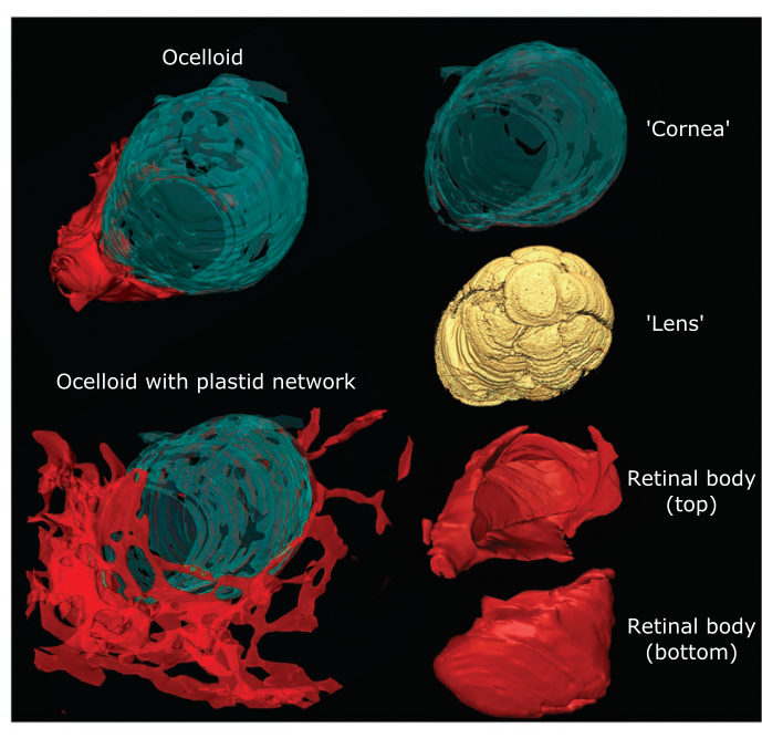
We mentioned that the equivalent of our cornea is made in Warnowiid by repurposing the endosymbiont mitochondria, show in green above. Similarly, the equivalent of our light-sensing retina is built in the Warnowiid using the photosynthesizing endosymbiotic plastid, shown in red in the lower right of the figure by Gavelis et al.
But check out in the lower left of the same figure — the plastid also shows up as an incredibly complex network that extends far beyond the retinal body. Why are these plastids forming a network? Is it possible that within the Warnowiid cell, these sub-cellular plastids are the equivalent of a neural net? Is the light signal captured by the plastid retinal body, somehow transferred to the plastid network, and processed? Do the plastids somehow convey that information to other organelles, the flagella, the piston, etc.? Does that network somehow control the hunting behavior of these already complex Warnowiids? These photosynthesizing, predatory algae with eyes?
Any way you look at them, these Warnowiids have a complexity that belies their humble single-celled status.
So let’s review again:
- First, over a billion years ago a bacteria-like cell learned to eat other cells.
- Then these single-celled eaters engulfed then re-purposed a spirochete-like bacterial cell to be its motor, the flagella.
- It has engulfed and re-purposed an aerobic bacteria-like cell to be its power plant, the mitochondria.
- It has engulfed and re-purposed a photosynthesizing bacteria-like cell to be its solar-energy-harvesting plastid.
- It may have engulfed and re-purposed another prokaryote to become its nucleus (though Margulis did not think so).
- And the ancestor to Warnowiids then re-purposed these now endosymbiotic organelles to build its eye.
We can see how that humble first step, the novel process of eating another cell, opened up an immense evolutionary frontier of complexity. But the complexity is not limited to big-brained animals like us. We see an amazing complexity even in single-celled organisms like the Warnowiid.
As Charles Darwin said in the closing sentence of On the Origin of Species:
“…whilst this planet has gone circling on according to the fixed law of gravity, from so simple a beginning endless forms most beautiful and most wonderful have been, and are being evolved.”