
#30DAYSOFSCIKUCHALLENGE
Cell-kus
A few cell-ebratory haikus and their deconvolution
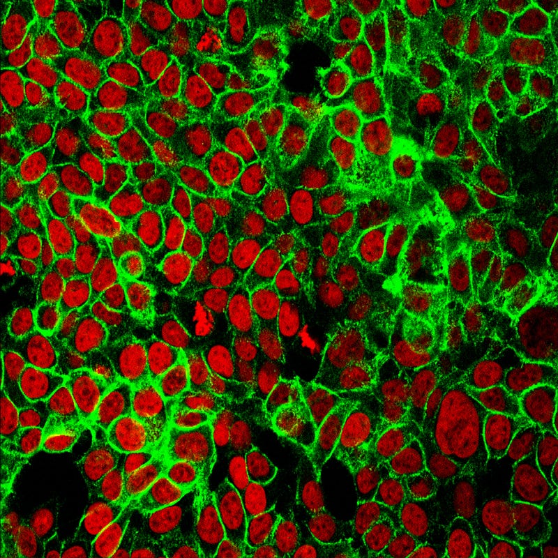
Cell Theory
The atoms of Life Building all Organisms Cells birthing more cells
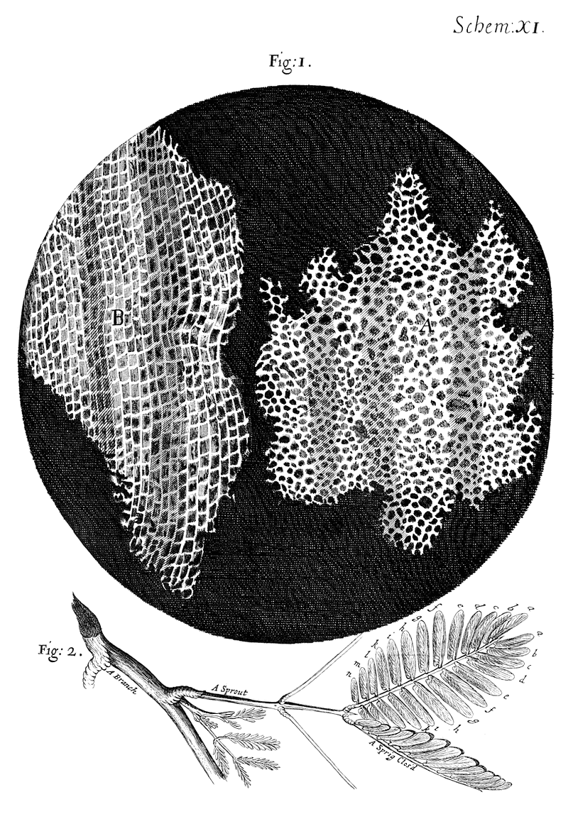
Robert Hooke, a polymath English scientist, published the first description of a micro-organism in his 1665 book, Micrographia. Hooke, along with Anton van Leeuwenhoek, a Dutch draper and self-taught builder of the most powerful microscopes of his time, introduced the world to a universe of microscopic life. Hooke’s drawing of a slice of cork showed the tiny compartments that reminded him of a monk’s room, or cell. Though Hooke gave cells their name, he did not originally think them alive. Leeuwenhoek was the first to propose that the microscopic organisms he saw, vigorously moving within their universe of a drop of water, were indeed living “animalcules.” Hooke confirmed the Dutchman’s observations, and together they pushed the boundaries of biology. To this day, microscopy in all its varieties anchors the discipline of cell biology.
In 1839 Jan Purkinje, Theodor Schwann, and Matthias Schleiden (and then Rudolph Virchow in 1855) proposed the earliest components of Cell Theory: that all living things are made of one or more cells; the cell is the basic unit of life, and all cells come from other cells.
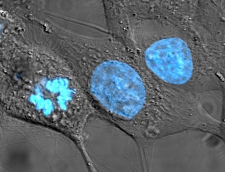
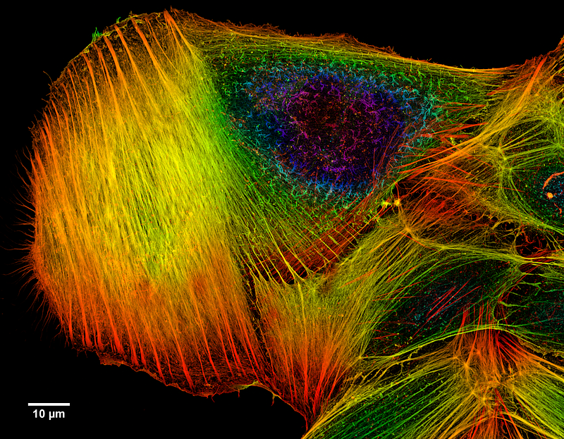
Many of the structures within cells are hard to resolve by unaided light microscopy. The explosion (sometimes literally) of organic chemistry in the late 1800s gave a tremendous boost to biology through the discovery of synthetic dyes. Microscopists discovered that certain dyes concentrated in certain regions of the cell, thus helping to dissect cell structure and function. Hoechst blue fluorescent nuclear dye shown in the bottom image is made by a German company (now wholly owned by a French company Sanofi), which was originally founded in 1863 as a chemical company and was one of the pioneers in biological stains.
The Kernel
Eukaryotic… cells harbor a nucleus Library of Life
Biologists separate cells into two broad categories. Prokaryotes are cells without a nucleus (pro meaning before, and karyon meaning kernel, or nucleus) and include all bacteria and a less familiar group called archaea. Bacteria and archaea arose billions of years before the nucleus. Eukaryotes are cells with a nucleus and include various single-celled organisms such as yeast and all the multicellular animals up to humans.
The nucleus is an organelle (a specialized structure within a cell), bounded by a membrane much like the cell’s own but doubled. The nuclear membrane separates the nuclear contents (the DNA containing the code for making the proteins the cell needs) from the rest of the cell, called the cytoplasm.
The evolutionary origins of the nucleus are shrouded in mystery, but fascinating theories abound. One theory suggests that the nucleus evolved from a virus and is shared here.
The cytoplasm is where most of the cell functions occur, one of the most important being to make the various proteins the cell needs. But the code for making proteins reside within the nucleus, separated by not one but two membranes, from the cytoplasm. So how does this work?
Within the nucleus, the code stored with the stacks of the DNA library is first copied into a message, a string of similar chemical composition called a messenger RNA or mRNA. This is the same type of chemical used in the latest vaccine against COVID-19, discussed here. The mRNA is transported out of the nucleus through nuclear pores guarded by protein-machines, which actively guard the nucleus and selectively shuttles specific molecules in and out only if they have the correct chemical password.
The mRNA then moves to the correct location in the cytoplasm, where it docks with a massive molecular machine called a ribosome. The ribosome reads the mRNA code and manufactures the protein per the instructions contained within. I discuss some of the core functions of the ribosomes in a little more detail in this article, under the section called “Saying goodbye to LUCA” — but of course, I think the whole article is worth a read, as it talks about the evolutionary origins of cells and Life. Enjoy!
Focus
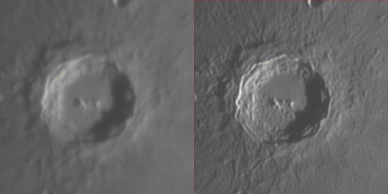
Deconvolution… brings focus to an image by removing noise
Microscopy and the practical physics of optics are exploding today. The ability to physically and computationally manipulate light brings incredible new abilities to focus microscopic images far beyond the normal physical limits of magnification as classically defined by optics (the Abbe limit of microscopic resolution from 1863). The technology can be applied to all images, not just microscopy — but the resolution on microscopy is stunning, as can be seen below.
For more on #30DaysOfScikuChallenge
What to read next? How about reading “Saying goodbye to LUCA” ScienceDuuude


